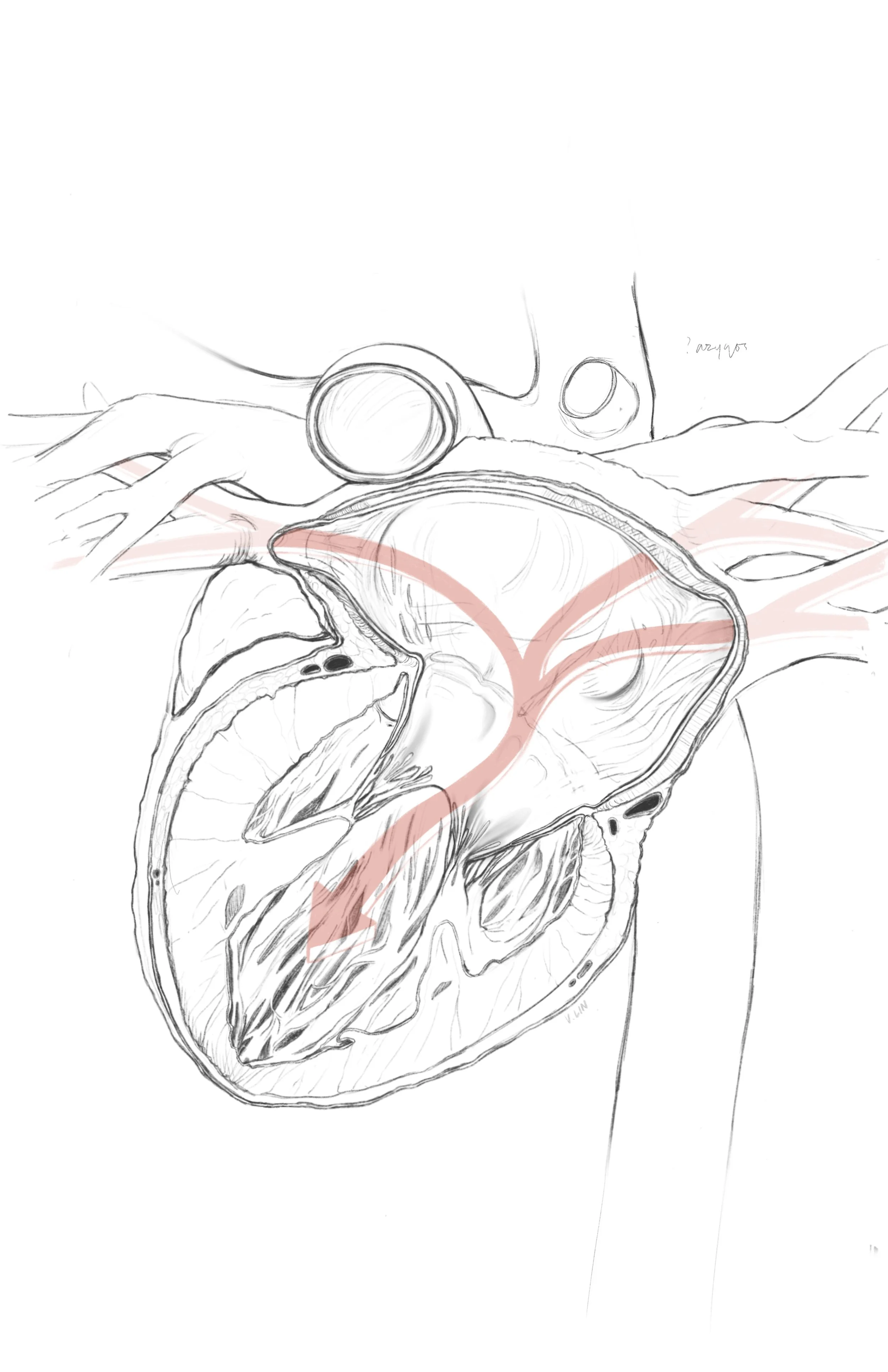
Posterolateral Inferior View of the Heart
2025
Digital Illustration
Created using Cinema 4D, Procreate, Adobe Photoshop, and Illustrator
Supervisor: Michael Corrin
Audience: General population
Format: Large-print educational poster
How do you show something that can’t really be seen?
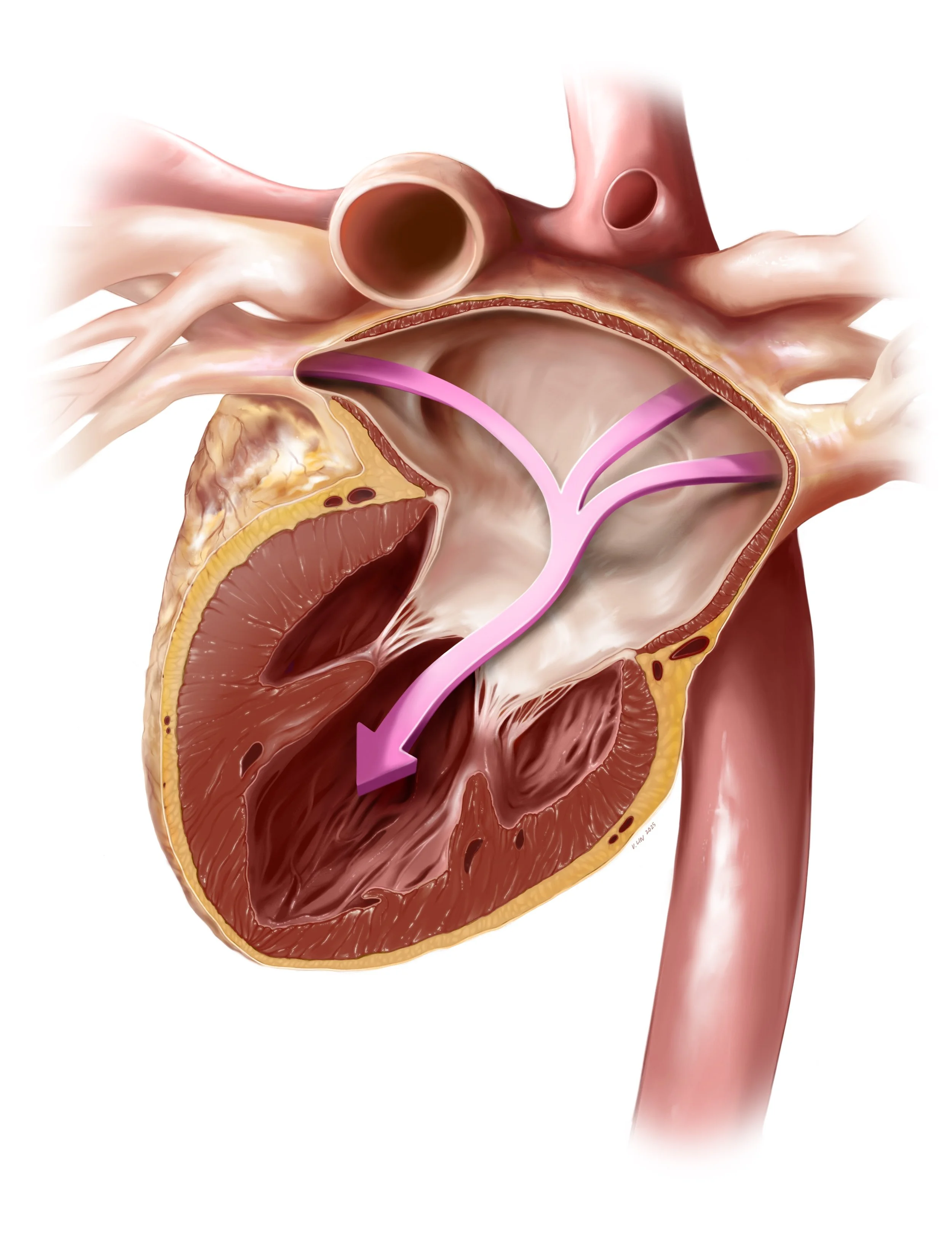
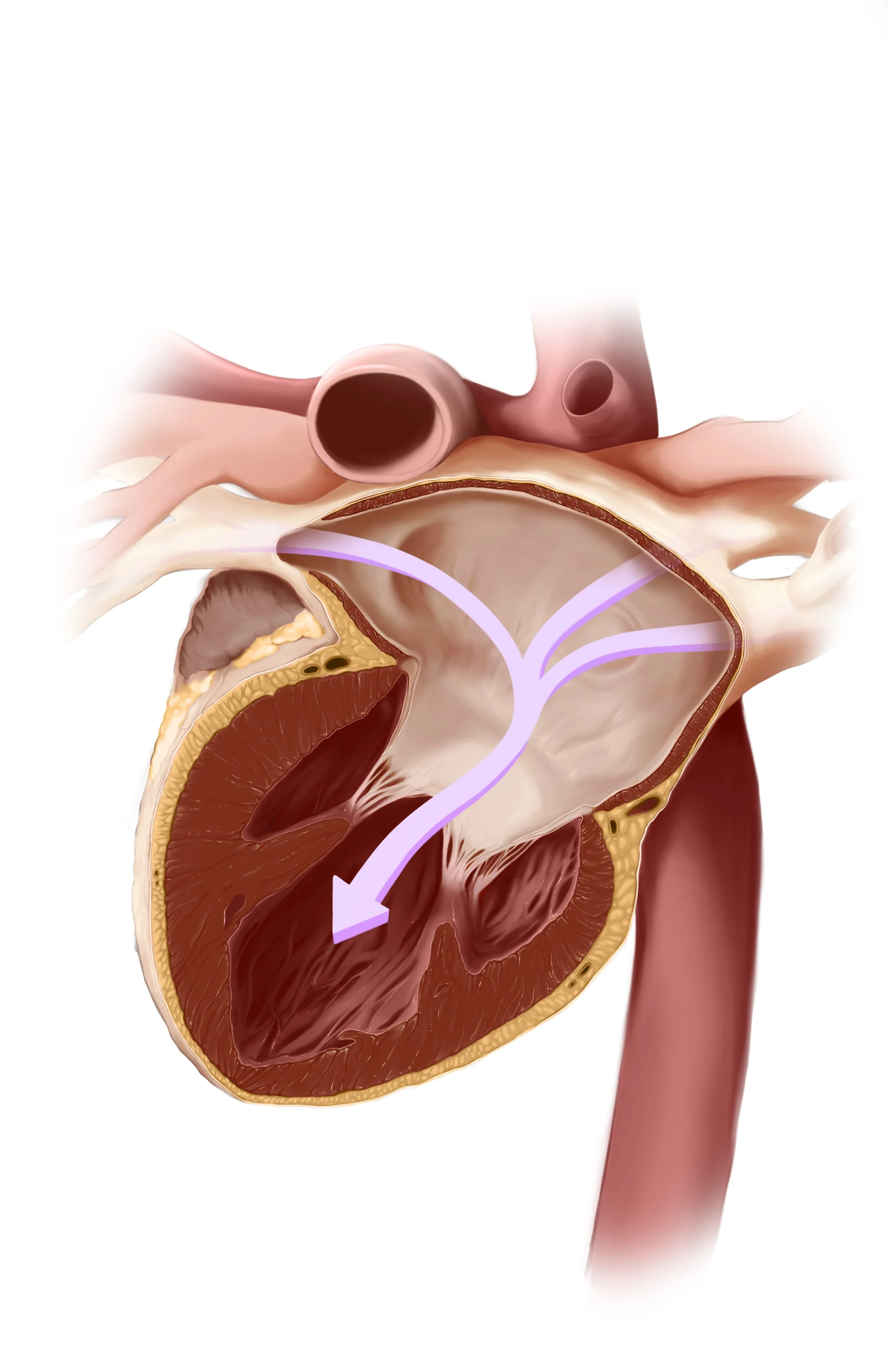
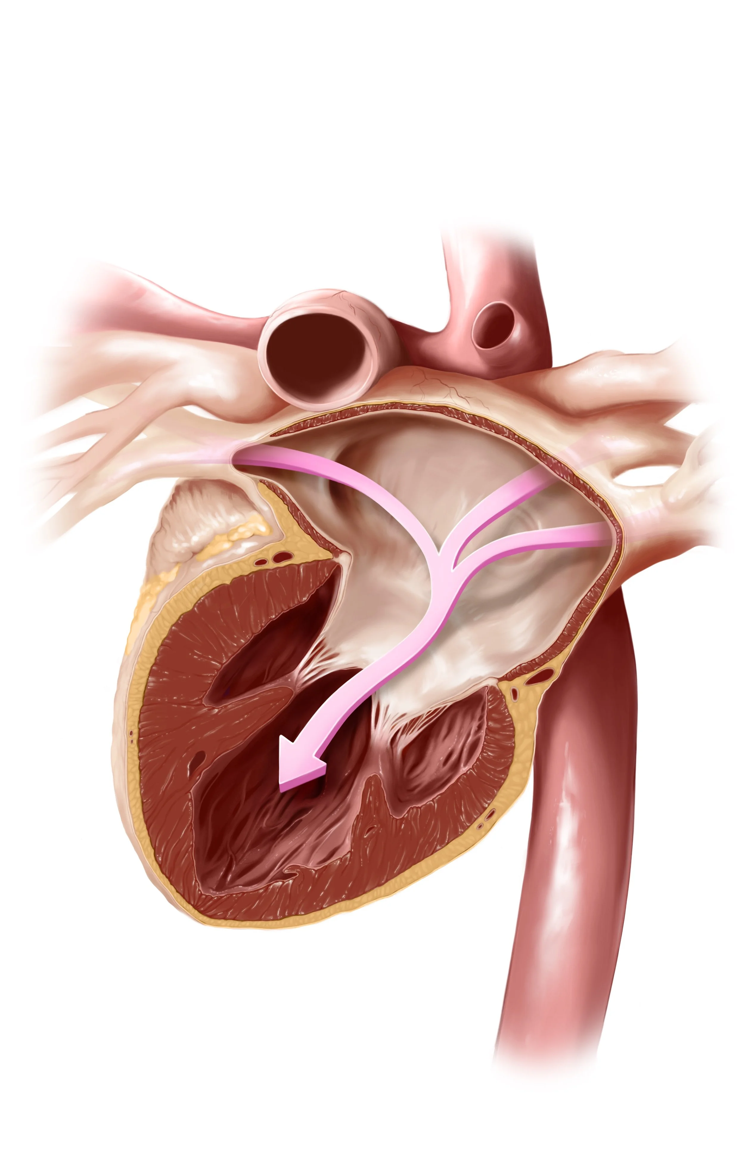
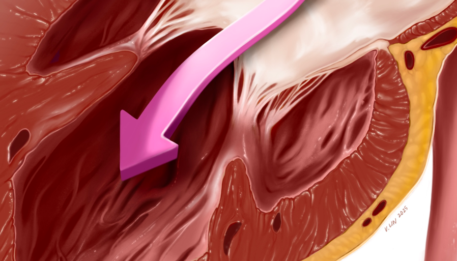
This was the challenge behind this illustration showcasing a posterior cross-section of the heart — specifically, the path of blood flow from the lungs into the left side of the heart (through the pulmonary veins, into the left atrium, and then the left ventricle).
In many educational illustrations of the heart, depictions rely on conventional perspectives (like frontal views) to showcase prominent features of heart anatomy. While conventional views can be helpful for this purpose, they often isolate the heart from its surrounding structures which limit our understanding of its spatial and functional complexity.
-
Understanding an anatomical system like the circulatory system requires familiarity with the broader context in which it exists, and depicting the heart in isolation can hinder one’s knowledge of its form, function, and relationships with closely-related organs like the lungs. This became the goal of this piece which aims to accurately portray the heart’s internal anatomy from a non-standard perspective while incorporating specific elements that situate the heart within its broader anatomical environment.
To fulfill this goal, I purposefully extended the visible major vessels on the exterior of the heart to emphasize how the heart connects to the lungs and fits within the thoracic cavity. By showing the branching of the pulmonary arteries and veins, the idea was to provide greater awareness to the underrated aspects of heart anatomy and give viewers a deeper appreciation for the organ in its living, intact form.
Illustration Process
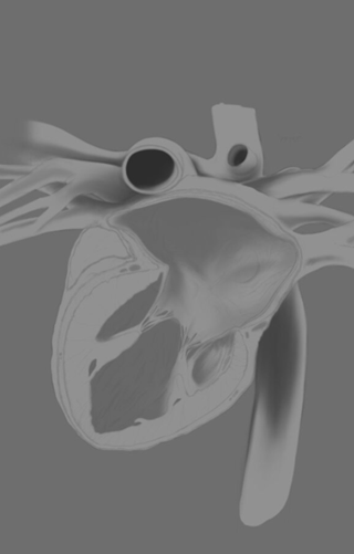
Research, Maquette-Building & Sketching
First, a 3D maquette was created in Cinema4D with the intended view and orientation of the cut plane. (Source: BodyParts3D)
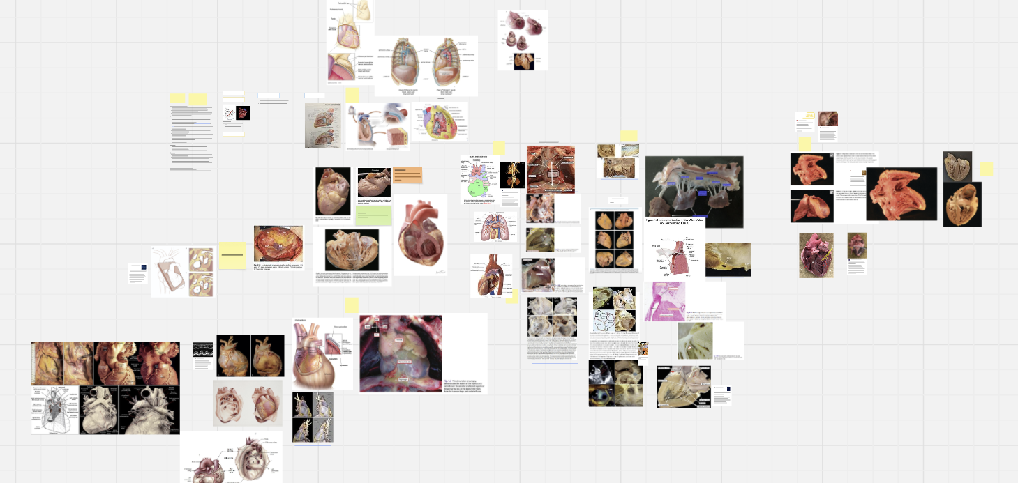
To develop the initial sketch for this piece, I dedicated extensive time researching heart anatomy from the posterior and mitral aspect. Sources included cardiac surgery textbooks and primary anatomical research literature.
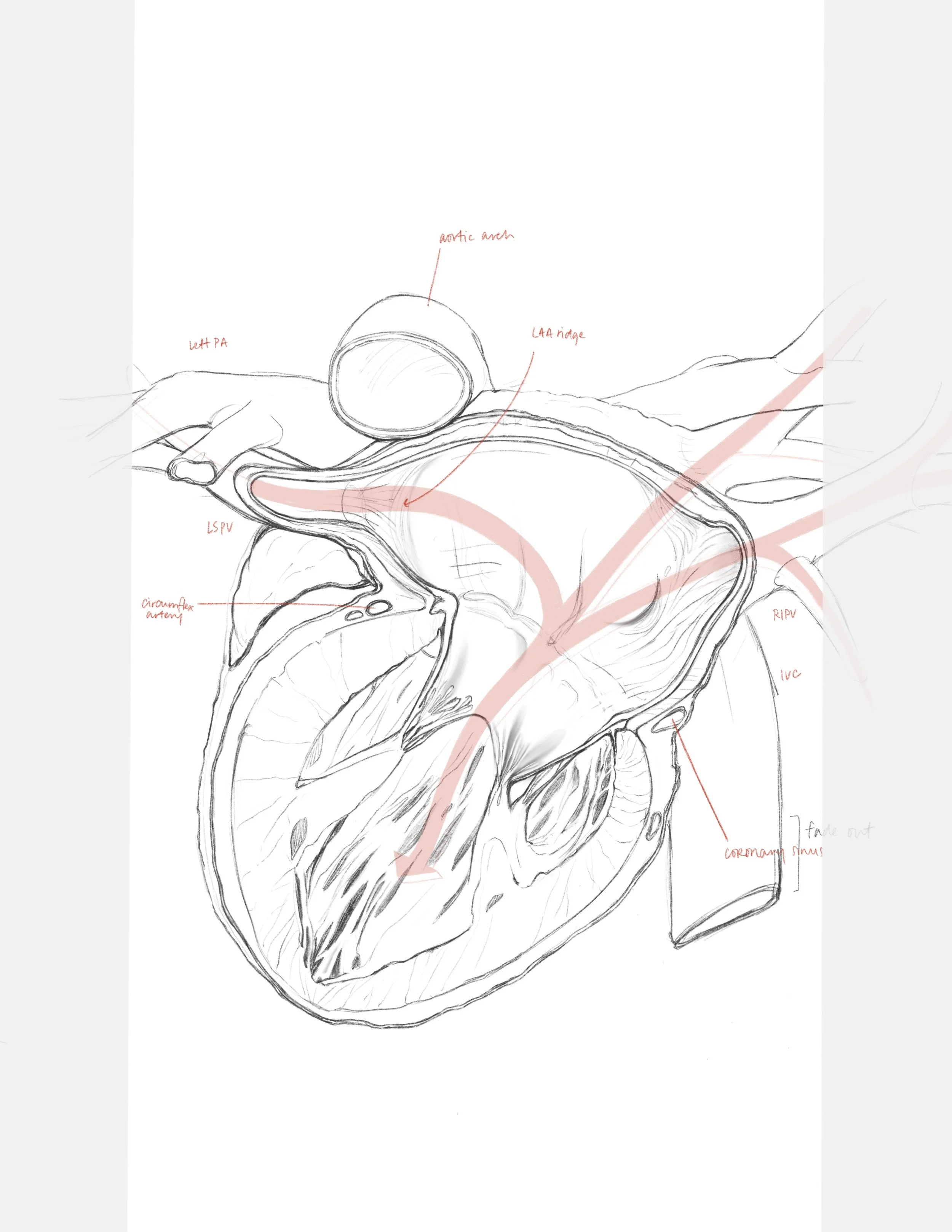
Then, I created the initial draft of the sketch and anatomical labels. The cut surface and internal textures were then refined with better detail.
For the final sketch, the truncated vessels were extended to illustrate the connections of the heart and emphasize the visual flow of the composition.
Greyscale Rendering
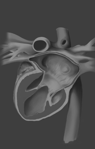
For the rendering portion, I opted to use the “Andrew Swift” rendering technique establish the full greyscale values of the piece. These tones would later become the basis for the colour values in the colourization stage.
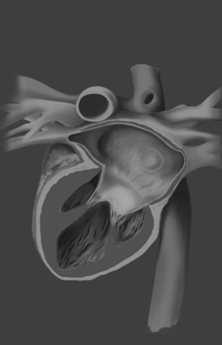
The interior of the left ventricle is rendered, paying close attention to the texture created by the trabeculated muscle fibers
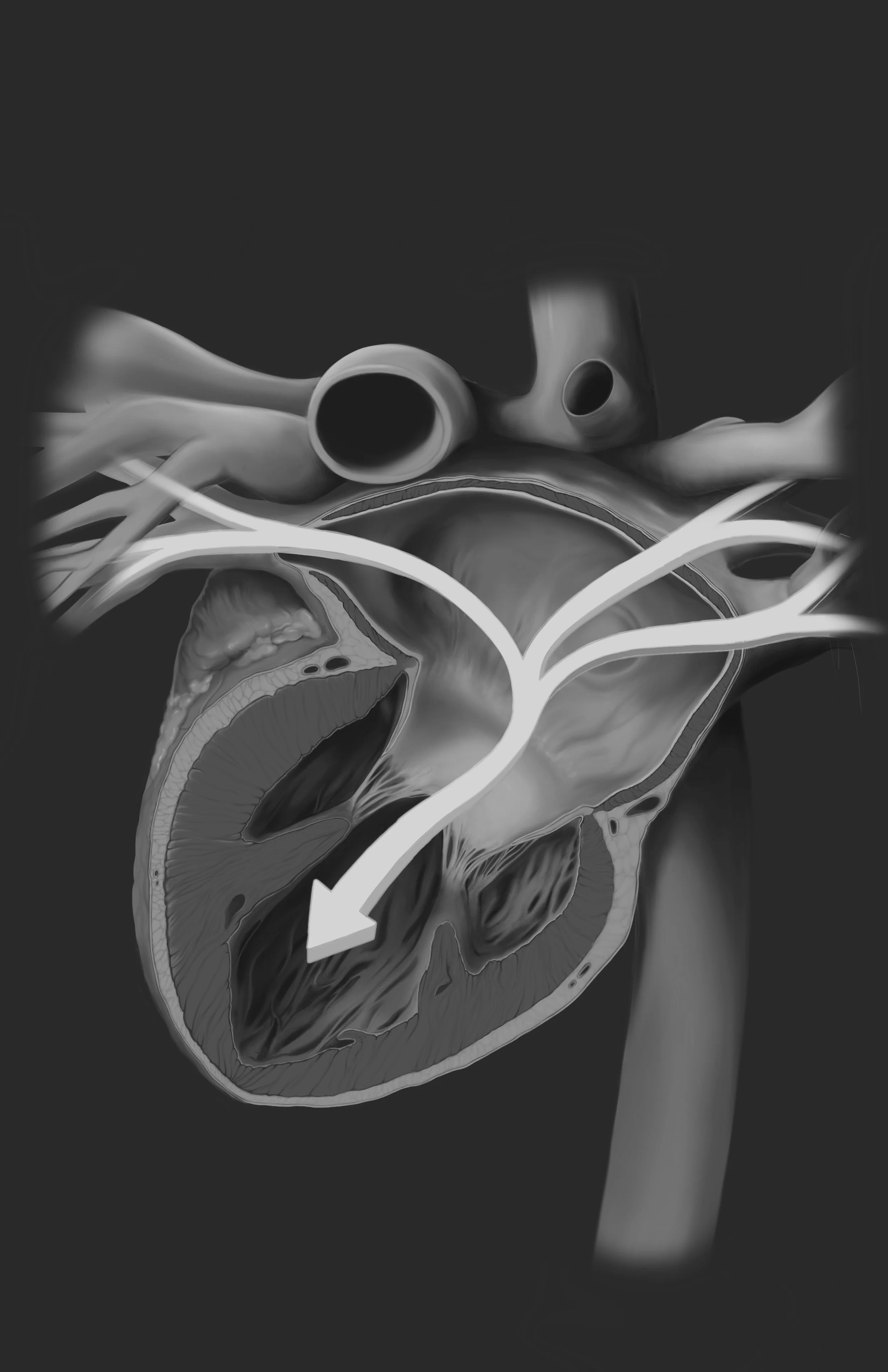
Continuing to push values, rendering the striations on the cut portion of the myocardium and adding highlights.
Colour Rendering
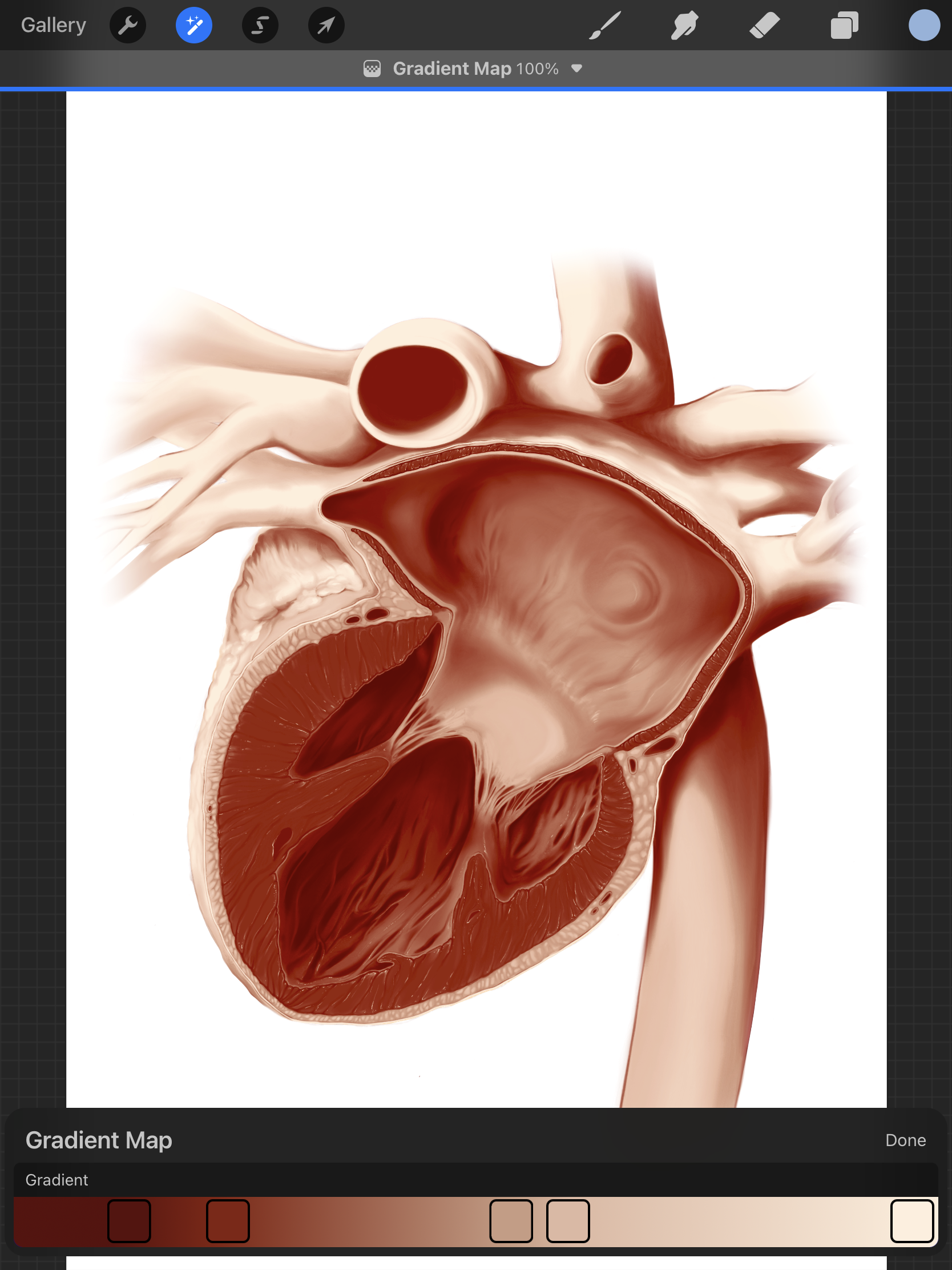
After the greyscale rendering was completed, a flesh-toned gradient map was applied to quickly add the base colours. Then, specific structures were isolated and overlaid with custom blending layers to create different hues that matched a normal heart specimen.
Next, the guiding arrows from the initial sketch were drawn in and different colours were tested to achieve optimal contrast.
Diffuse and specular highlights were added to evoke the sense of living tissue.
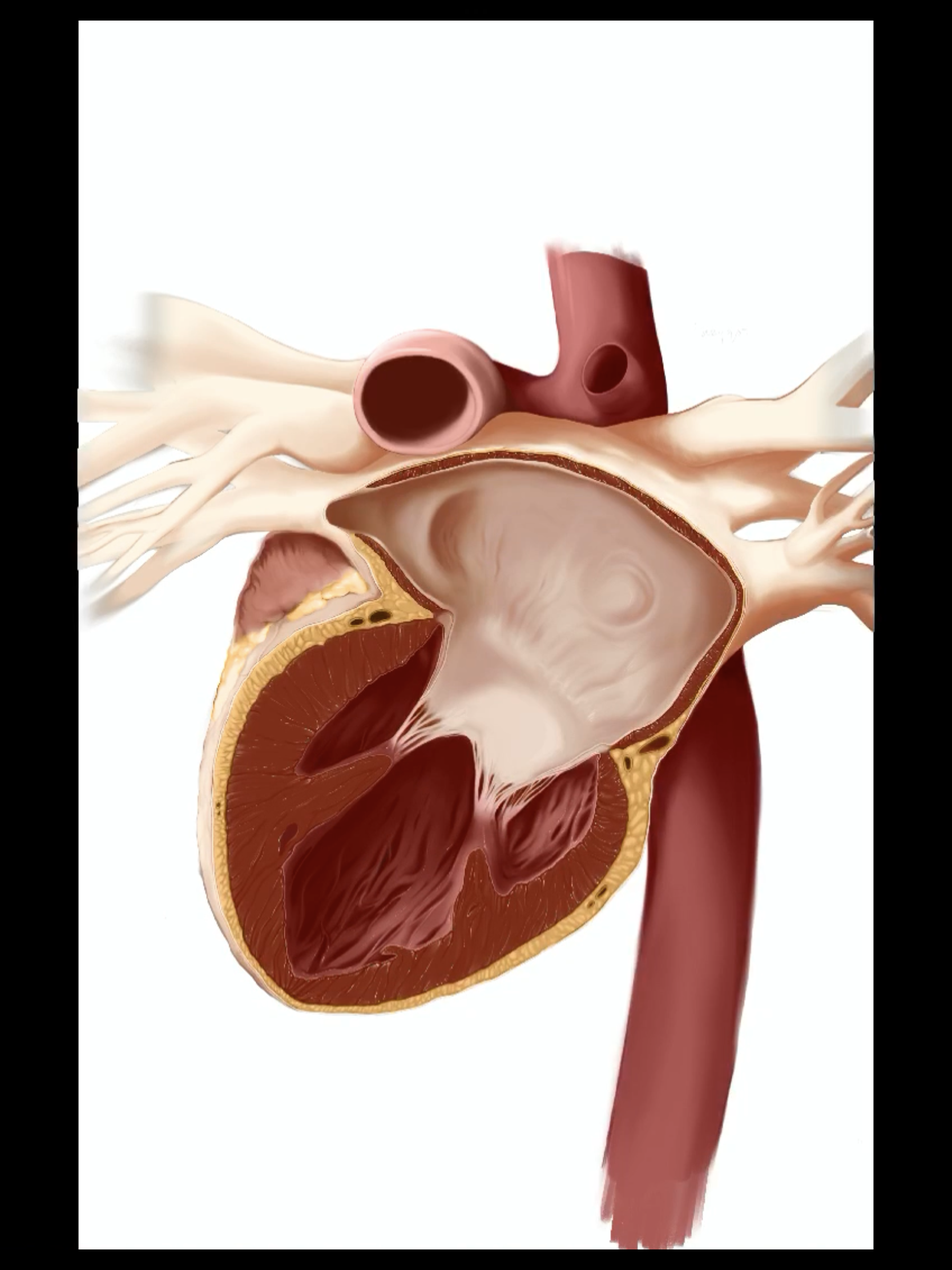
Revisions
After the initial completion of the illustration, it became clear that more could be done to push the realism and life-like qualities of this piece and present the flesh in a more accurate way.
This included refining the following aspects:
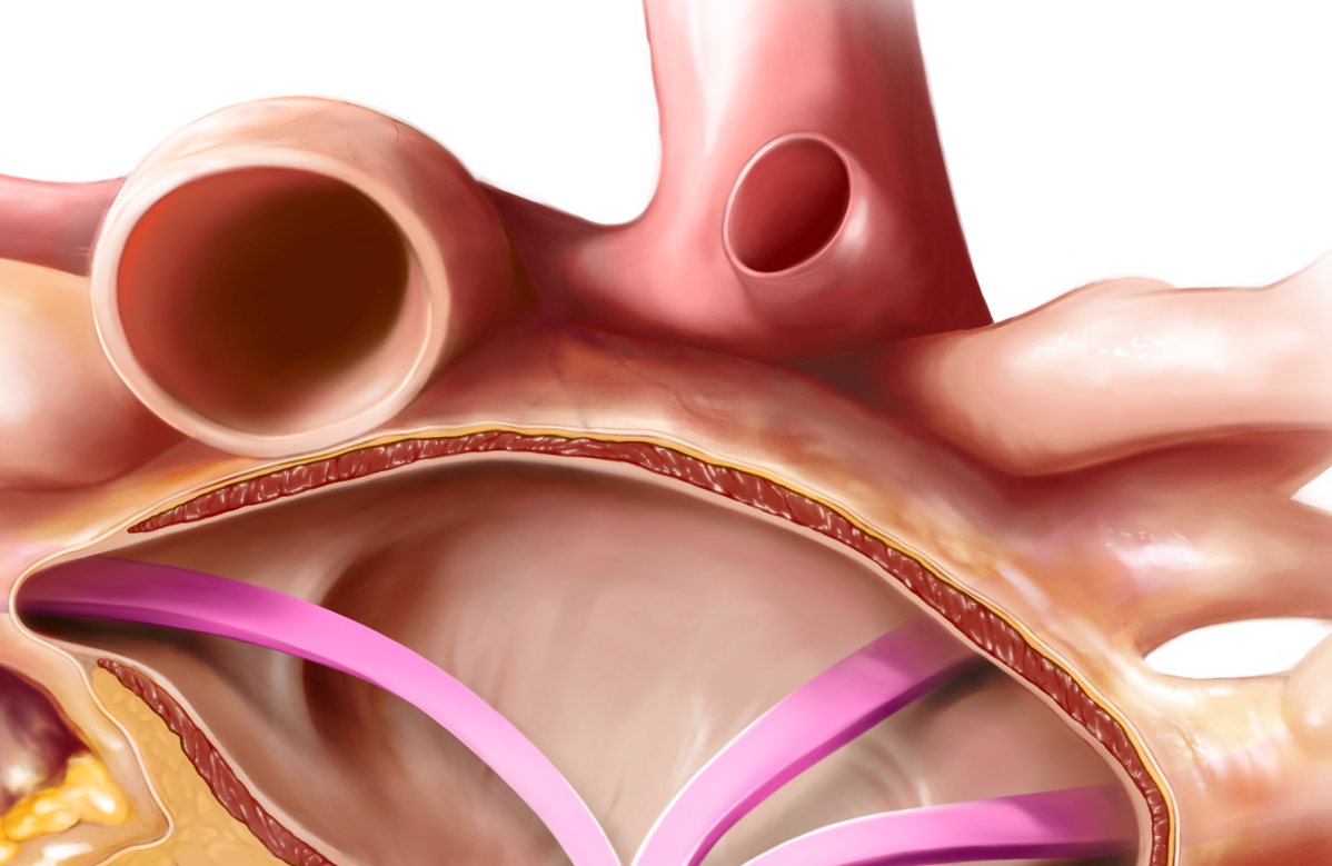
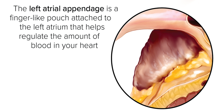
Revising the pericardium surrounding the left atrial appendage to appear more continuous around the heart
Adding cast shadows falling onto the arrow to appear more tangible or integrated in the scene
Adding subsurface scattering details (to simulate translucency) and microvasculature textures
Bringing Everything Together
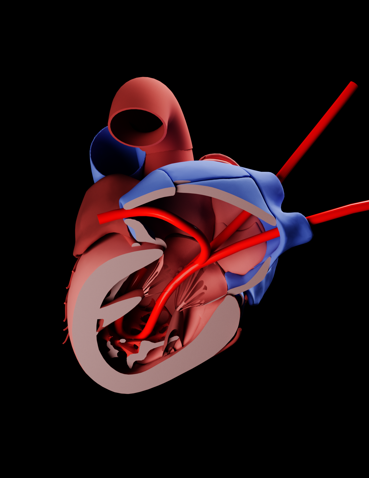
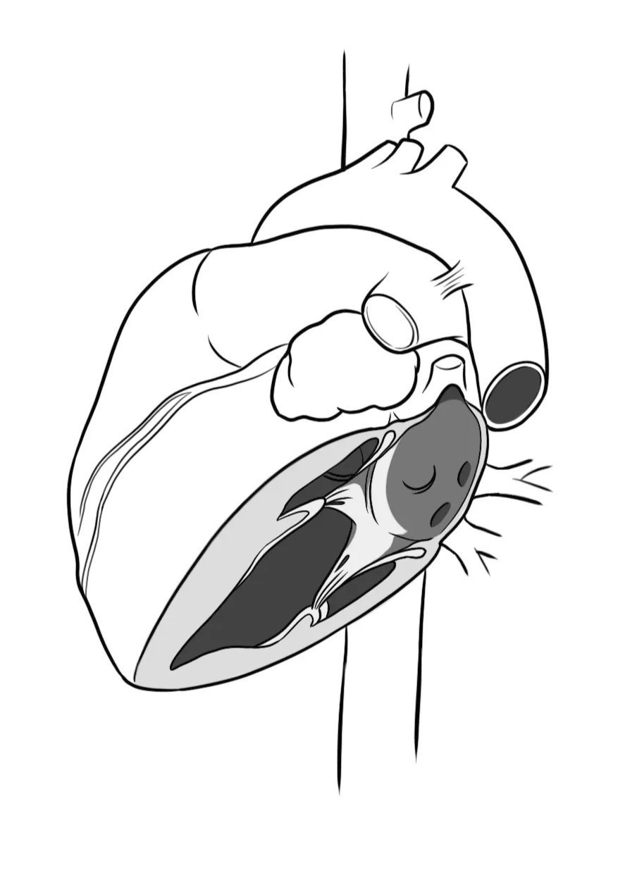
To tie everything together into a cohesive, didactic-style piece, I included supplementary visual aids to help viewers better conceptualize the heart’s orientation within its spatial environment. Specifically, I created a simplified schematic diagram of the illustrated heart from a lateral view to clarify its positioning relative to the body.
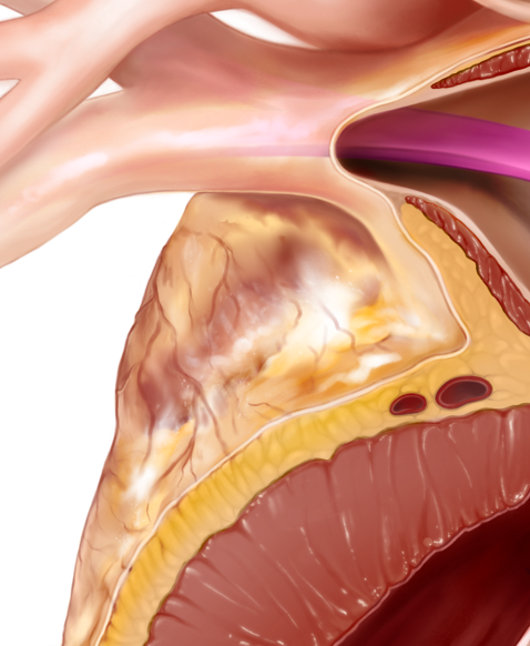
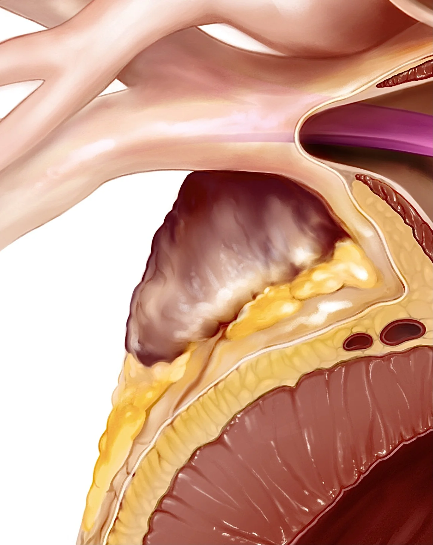
Additionally, I chose to prominently feature the left atrial appendage in the final poster. Its inclusion was both a visual and conceptual decision, given its significance in the anatomy of the left atrium and its relevance in the context of blood flow.
Conclusion
Overall, this project challenged me to think beyond conventional anatomical views and explore new ways of visualizing internal structures that are typically hidden from sight. Balancing anatomical accuracy with visual storytelling required continuous iteration, research, and design problem-solving.
Through this process, I deepened my understanding of spatial anatomy and gained valuable experience in conveying complex biological concepts through illustration. Ultimately, this piece represents not only a study of the heart’s structure and function, but also my commitment to creating visuals that inform, engage, and inspire curiosity in both clinical and educational settings.
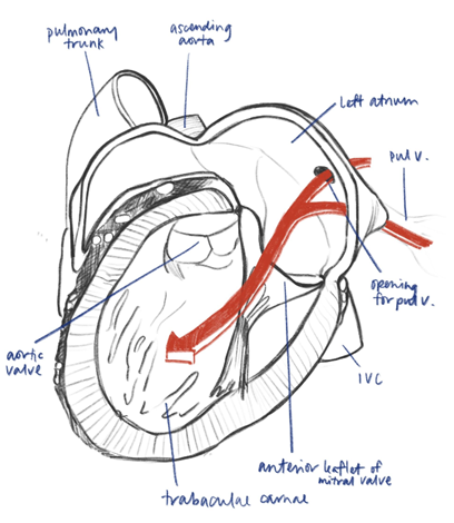
Initial draft iteration of this piece