
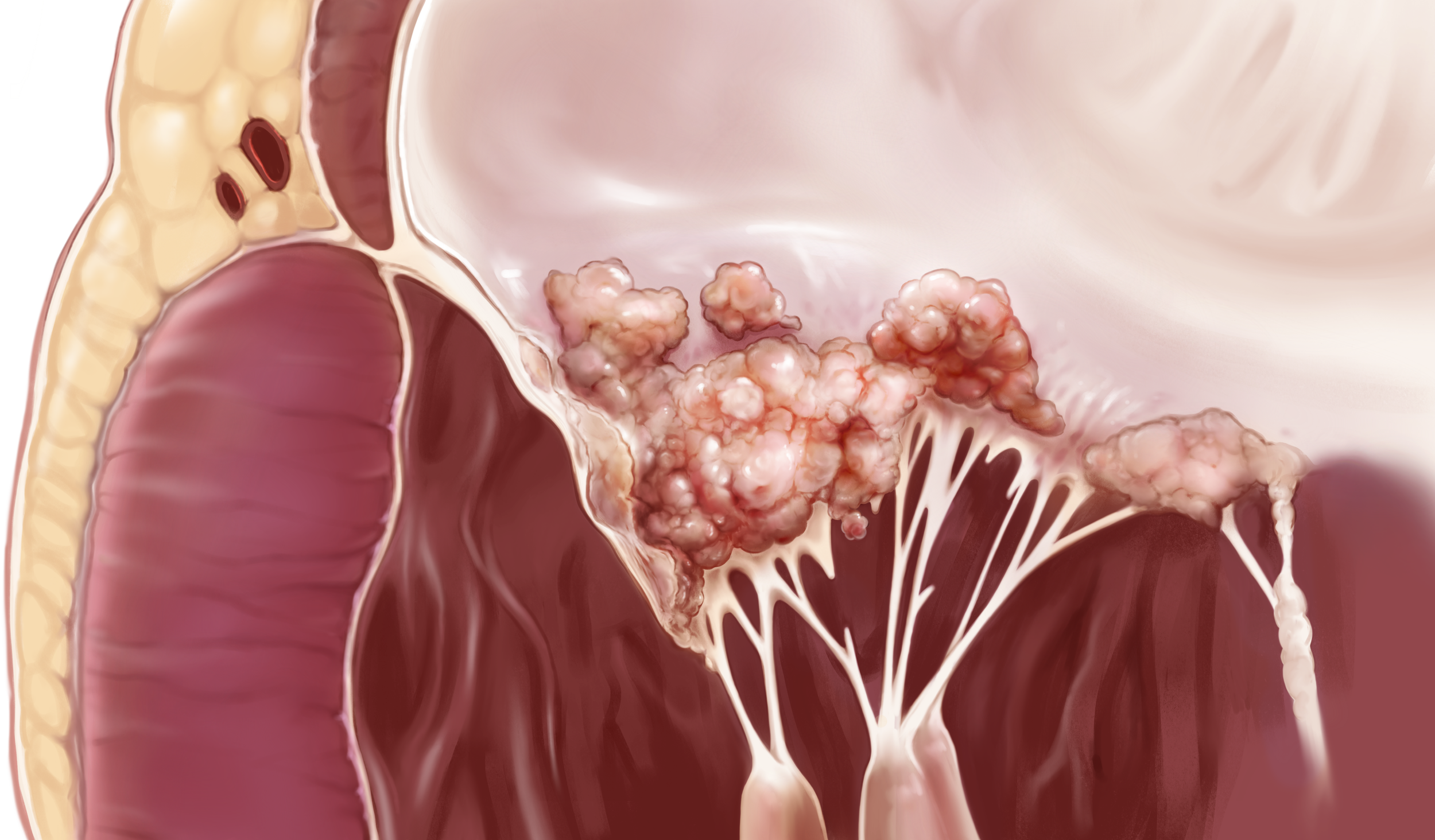
Infective Endocarditis
2026
Pathology Infographic
Created using Adobe Illustrator, Photoshop, Procreate
Supervisor: Dave Mazierski, Dr. John Wong
Audience: General scientific audience
Format: 2-page magazine spread
Project Description
Visualizing the Pathophysiology of Infective Endocarditis
Infective Endocarditis (IE) is a rare, but deadly infection that affects the inner lining of the heart called the endocardium. This two-page magazine spread illustrates the progression of this disease by highlighting the key players and events that facilitate the colonization, proliferation and eventual destruction of heart valves if left untreated.
Jump To:
I. Research & References
I. Tissue Studies
II. Illustrating Insets & Secondary Graphics
III. Graphic Design
Researching the Pathophysiology of IE
The development of IE carries implications across the molecular, cellular, tissue, and system level. Factors at each level can be hijacked when an infection occurs which signals the breadth of visual approaches to explain the development of this pathology. While researching the literature surrounding this topic, I became fascinated by how pathogens like Staphylococcus aureus hijack normal cellular responses to injury and healing to create the perfect storm of events that sustain its colonization.
Thus, I wanted to craft the narrative of this pathophysiology by not only explaining the cellular events upon infection, but also how these effects compound over time — leading up to the consequences you see on the tissue and systemic scale.
Meanwhile, I collected reference photos to study the unique textures and appearances of heart tissue, as well as images of diseased tissue.
Tissue Studies
Insets & Secondary Graphics Process
Progression of IE Insets
Initial sketch detailing the three key stages of the pathophysiology progression, showing the main cell types involved. These include activated platelets, lymphocytes, endothelial cells, as well as fibrin proteins to illustrate the coagulation process.
Refined sketch of the first stage of the pathophysiology
The sketch was simplified for clarity before it was colourized
Later, I went back and changed the colour of the blue platelets to make it look more natural